Wednesday, May 27, 2009

Tufted Bacteria Cause Infection In Premature Babies

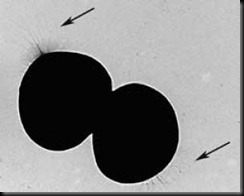
Bacteria that normally reside on the skin of healthy people can cause serious infections in premature babies. A group of researchers at the Swedish medical university Karolinska Institutet have now found an explanation for why a certain kind of staphylococcus can attach itself to the skin and quickly develop dynamic ecosystems: the bacteria are like tufted, self adhesive hairballs.

Staphylococcus establishes itself on the child's skin and mucous membranes directly after birth. In healthy adults and children, these bacteria normally live in harmony with the host organism. However, in sick adults or premature babies, they can cause blood poisoning.

Tufted  bacteria

The scientists believe that the hair-like protrusions on the surface of the bacteria that have now been identified serve to adhere the bacteria to the host's cells, whereupon they cause infection. They also found that the antimicrobial substance LL37, which is found on the skin (amongst other places) can inhibit the growth of the bacteria, and probably plays an important part in keeping the bacteria flora stable and inhibiting their uncontrolled proliferation.

"We wanted to conduct this research not only to learn more about the pathogenic potential of the bacteria, but also to understand how the child can protect itself from attack by, for instance, enhancing the body's own defences," says Giovanna Marchini, associate professor at Karolinska Institutet and senior physician at the Astrid Lindgren Children's Hospital neonatal section.

Dr Marchini stresses that humans have evolved effective forms of co-existence with certain microbes; for example, the most common intestinal bacteria produces Vitamin K, which we need every day and which is important for the blood's coagulative properties. Bacteria are also necessary for the development of an effective immune defence system. In recent years, these 'beneficial' bacteria have been the object of increasingly intensive study, and are behind the development of the 'hygiene theory'.

"It's thought that the past decades' hunt for disease-causing bacteria means that we now live too cleanly, which has contributed to the sharp rise in allergies and other ‘luxury diseases'," continues Dr Marchini. "Other than wanting to prevent infection in babies, we also think it's an exciting challenge to understand the conceivable health aspects of these tiny, round and tufted skin dwellers.

Source: Sciencedaily
Monday, May 18, 2009

Acne antibiotic may be treatment for brain fever

Minocycline reduces death in JE-infected mice and could do the same in humans, say researchers.If Indian scientists can transfer their laboratory success to human trials, a common acne-treating antibiotic could well turn out to be a life saver for patients of Japanese encephalitis (JE), commonly known as brain fever.

Researchers at the National Brain Research Centre (NBRC) in Manesar, Haryana, have shown that the common antibiotic minocycline reduces death in JE-infected mice and believe it could do the same in humans.

Pill_Bottle

“Even if we replicate a fraction of the success in animals reported by NBRC team, we can save several lives and prevent many others from being maimed every year,” says N.C. Borah, managing director of the Guwahati Neurological Research Centre (GNRC).

He plans to undertake clinical trials in May at GNRC and two other hospitals in Guwahati and Dibrugarh as each of these centres receive 300-400 seriously ill JE patients every season that typically begins with the monsoon.

A viral disease that causes inflammation of the brain, JE kills about one-third of its patients and half the remaining end up with serious disabilities including cognitive and behavioural impairment. The infection is endemic in India, where 135 districts in 15 states routinely report cases, according to the Union ministry of health and family welfare.

“No effective treatment exists today, what we have is only supportive,” says Anirban Basu, the lead researcher from NBRC, who thinks clinical success could lead to immediate therapy, as minocycline is an inexpensive and freely available drug currently used for treating bacterial infections of the skin, teeth and urinary tract.

Basu’s early work showed that minocycline not only reduced the inflammation in the brain, but also stemmed loss of healthy neuronal cells and decreased the viral load (a measure of the severity of a viral infection). However, questions like how effective the drug will be, if there’s a time lag between the infection and the treatment, or whether it will reduce the disabilities in survivors can be answered only after the human trials, says Basu.

Scientists believe it’s a fairly safe bet, as it’s the efficacy rather than the safety that needs to be ascertained, because the drug is already in use. “It’s a straightforward case, but we are hitting a stone wall,” says NBRC director Vijayalakshmi Ravindranath, since there’s no government agency to do translational research or even coordinate it. Translational research helps convert laboratory findings into therapeutics—or from bench to bedside. “In the absence of intellectual property, there’s no money to be made here or else we could have gone to a commercial agency for trials.”

The cost of the trial is not a trivial issue either. But the department of biotechnology, of which NBRC is an autonomous institute, has promised to extend support. Trials in children are difficult and expensive, as each centre needs to have sufficient number of ventilators, which can cost up to Rs 20,000 per day for every child. “I am pushing it; we hope to get started by this season,” says Ravindranath. NBRC has identified a contract research organization and is chalking out the trial design, things which research institutions in the country typically don’t do.

Borah thinks the sample of 250 patients can be recruited in just one season in the north-east, which has the highest incidence of JE. “We will run the study for four weeks and it can be completed by November,” he adds.

The successful and timely completion of this trial is important for more than one reason. Minocycline has shown promising results in other neurogenerative disorders including Alzheimer’s and Parkinson’s disease and multiple sclerosis, a nervous system disorder. Moreover, its success in JE could be an encouragement for further initiatives in “repurposing” of existing or discarded drugs. “This is a good strategy for developing countries,” says Basu.

Source:TOI

Friday, May 15, 2009

9 Foods That Reduce Stress Levels


Eat these foods, reduce your stress.

Reach for these items next time you're feeling under pressure, under the weather, or just too close to that breaking point. Munching on these stress-free foods will help pull you back into the game.

                                                       Oranges

oranges

A German study in Psychopharmacology found that vitamin C helps reduce stress and return blood pressure and cortisol to normal levels after a stressful situation. Vitamin C is also well-known for boosting your immune system.

                                              Sweet Potatoes

Sweet-Potato

Sweet potatoes can be particularly stress-reducing because they can satisfy the urge you get for carbohydrates and sweets when you are under a great deal of stress. They are packed full of beta-carotene and other vitamins, and the fiber helps your body to process the carbohydrates in a slow and steady manner.

                                                   Dried Apricots

dried apricot

Apricots are rich in magnesium, which is a stress-buster and a natural muscle relaxant as well. Almonds, Pistachios & Walnuts. Almonds are packed with B and E vitamins, which help boost your immune system, and walnuts and pistachios help lower blood pressure.

                                                      Turkey

turkey Turkey contains an amino acid called L-tryptophan. This amino acid triggers the release of serotonin, which is a feel-good brain chemical. This is the reason why many people who eat turkey feel relaxed, or even tired, after eating it. L-Tryptophan has a documented calming effect.

                                                    Spinach

spinach A deficiency in magnesium can cause migraine headaches and a feeling of fatigue. One cup of spinach provides 40 percent of your daily needs for magnesium.

                                                   Salmon

salmon Diets high in omega-3 fatty acids protect against heart disease. A study from Diabetes & Metabolism found that omega-3s keep the stress hormones cortisol and adrenaline from peaking.

                                                   Avocados

avocado The monounsaturated fats and potassium in avocados help lower blood pressure. The National Heart, Lung, and Blood Institute says that one of the best ways to lower blood pressure is to consume enough potassium (avocados have more than bananas).

                                            Green Vegetables

broccoli Broccoli, kale, and other dark green vegetables are powerhouses of vitamins that help replenish our bodies in times of stress.

More stress-busting tips:

  • Exercise reguarly
  • Drink an energy shake for breakfast
  • Eat small meals throughout the day, which will keep your blood sugar stable (when blood sugar is low, mental, physical, and emotional energy decreases, and stress increases).

Source:Msn

Wednesday, May 13, 2009

Bacteria Play Role In Preventing Spread Of Malaria

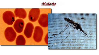
Bacteria in the gut of the Anopheles gambiae mosquito inhibit infection of the insect with Plasmodium falciparum, the parasite that causes malaria in humans, according to researchers at the Johns Hopkins Bloomberg School of Public Health.

Scientists found that removing these bacteria, or microbial flora, with antibiotics made the mosquitoes more susceptible to Plasmodium infection because of a lack of immune stimulation. As part of the malaria transmission cycle, a mosquito acquires the malaria-causing parasite when it feeds on blood from an infected person. The parasite develops within the mosquito and can then be transmitted to another human when the mosquito feeds again.

malaria_pics

“Our study suggests that the microbial flora of mosquitoes is stimulating immune activity that protects the mosquito from Plasmodium infection. The same immune factors that are needed to control the mosquito’s infection from the microbes are also defending against the malaria parasite Plasmodium,” said George Dimopoulos, PhD, senior author of the study and associate professor with Johns Hopkins Malaria Research Institute.

“The interplay between bacteria and the mosquito’s immune system may have significant implications for the transmission of malaria in the field where mosquitoes may be exposed to different types of bacteria in different regions. Theoretically, these bacteria could be introduced to the mosquitoes to boost their immunity to the malaria parasite and make them resistant and incapable of spreading the disease. Our current research aims at identifying those bacteria that trigger the strongest mosquito immune defense against the malaria parasite.”

As part of the study, the Johns Hopkins researcher treated mosquitoes with antibiotics to kill the gut bacteria. Treated mosquitoes were more susceptible to infection by Plasmodium when feeding on infected blood compared to mosquitoes that were not treated with antibiotics. To further verify the results, bacteria-free mosquitoes were infected with bacteria to determine if they were less susceptible to Plasmodium infection.

In addition, the researchers determined that mosquitoes infected with bacteria died earlier than mosquitoes without bacteria when infected with Plasmodium; 60 percent of the mosquitoes with gut-bacteria died compared to 40 percent of those free of bacteria—even with Plasmodium levels five times higher than those with bacteria.

“The malaria parasite must live in the mosquito for about two weeks in order to complete its life cycle and be transmitted to a person. The fact that these bacteria shorten the mosquito’s life span is additional good news,” said Dimopoulos.

Malaria kills over one million people worldwide each year; the majority of deaths are among children living in Africa.

Source:Sciencedaily

Friday, May 8, 2009

Thalassemias

On 8th May Thalassemias Day is celebrated to make people aware about silent inherited disease which affecting younger population. There are around 30 crore carriers in India and every year 10,000 children with thalassemia major are born in India , which constitutes 10% of the total number in the world. It is common in Sindhis, Punjabis, Gujaratis, Lohana etc.

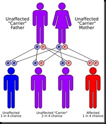
Thalassemias  are inherited blood disorders. "Inherited" means they're passed on from parents to children through genes. Thalassemias cause the body to make fewer healthy red blood cells and less hemoglobin  than normal.

Hemoglobin contains a large amount of iron. When red blood cells are broken down, most of the iron from the hemoglobin is used again to make new hemoglobin. In the case of thalassemia the hemoglobin is fragile and breaks down sooner than normal, thus leaving the person with not enough hemoglobin in their body. This lack of hemoglobin causes anemia.

Different Types of Thalassemia

  • Alpha (a) Thalassemia: Alpha thalassemia results in an excess of beta globins, which leads to the formation of beta globin aggregates called hemoglobin H. These aggregates are more stable and soluble, but under special circumstances can lead to hemolysis, generally shortening the life span of the red cell.
  • Beta (b) Thalassemia: Beta thalassemia is a thalassemia in which there is a decreased production of beta globin chains. The excess alpha chains aggregate and the degree to which these chains aggregate determines their severity.

Causes :

b183942897

Thalassemia is an inherited disease, which means it is passed on from parents to their child through their genes. It is an autosomal recessive trait, it is not linked by sex chromosomes. It is not infectious and cannot be “Caught” like a cold. It will not develop later in life, nor can a child outgrow it. Both parents must have thalassemia trait in order to pass the disease on to their child, but it only takes one parent to pass trait on to his/her child. Thalassemia trait will never develop into disease. Thalassemia trait can be passed on for many generations without being detected before a child is born with disease.

Symptoms:

People with thalassemia major may experience the following:

  • Paleness
  • Headaches
  • Fatigue
  • Shortness of breath
  • Jaundice
  • Spleen enlargement

Diagnosis:

The diagnosis of thalassemia trait and thalassemia major is made from microscopic examination of the blood, which shows many small, pale red blood cells, and from other blood tests that show reduced levels of adult hemoglobin in the blood. Diagnosis is confirmed by doing Hemoglobin electrophoresis on a sample of blood. The deficient synthesis of the hemoglobin are demonstrated by this test. In addition if the patient has hemolysis or breakdown of red blood cells there will be increased levels of bilirubin and fragmented cells on the peripheral blood smear for hemolysis.

Treatment:

Normally, there are no treatments recommended. However, the doctor may suggest taking iron medication if they feel it is necessary.

The primary treatment is regular blood transfusions, usually every four weeks. In addition to the blood transfusions, doctors recommend injections of Deferral to help the body flush out the extra iron created by the new blood. The injections are given under the skin from a small pump 5 to 7 nights a week.

Additionally, splenectomy (removal of the spleen), bone marrow transplants and chelation therapy are being researched as possible treatments for thalassemia.

Thursday, May 7, 2009

Microscopic Images

Get up close and personal with your innards with these 15 amazing 3D-body shots. Almost all of the following images were captured using a scanning electron microscope (SEM), a type of electron microscope that uses a beam of high-energy electrons to scan surfaces of images. The electron beam of the SEM interacts with atoms near or at the surface of the sample to be viewed, resulting in a very high-resolution, 3D-image. Magnification levels range from x 25 (about the same as a hand lens) to about x 250,000. Incredible details of 1 to 5 nm in size can be detected.

Max Knoll was the first person to create an SEM image of silicone steel in 1935; over the next 30 years, a number of scientists worked to further develop the instrument, and in 1965 the first SEM was delivered to DuPont by the Cambridge Instrument Company as the “Stereoscan.

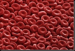
1. Red blood cells

RBC They look like little cinnamon candies here, but they’re actually the most common type of blood cell in the human body - red blood cells (RBCs). These biconcave-shaped cells have the tall task of carrying oxygen to our entire body; in women there are about 4 to 5 million RBCs per microliter (cubic millimeter) of blood and about 5 to 6 million in men. People who live at higher altitudes have even more RBCs because of the low oxygen levels in their environment.

2. Split end of human hair

Split human hair Regular trimmings to your hair and good conditioner should help to prevent this unsightly picture of a split end of a human hair.

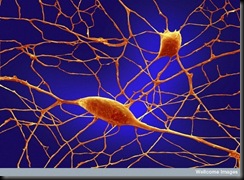
3. Purkinje neurons

neurons Of the 100 billion neurons in your brain, Purkinje neurons are some of the largest. Among other things, these cells are the masters of motor coordination in the cerebellar cortex. Toxic exposure such as alcohol and lithium, autoimmune diseases, genetic mutations including autism and neurodegenerative diseases can negatively affect human Purkinje cells.

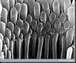
4. Hair cell in the ear

Hair cell in ear

Here’s what it looks like to see a close-up of human hair cell stereocilia inside the ear. These detect mechanical movement in response to sound vibrations.

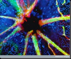
5. Blood vessels emerging from the optic nerve

optic nerves In this image, stained retinal blood vessels are shown to emerge from the black-colored optic disc. The optic disc is a blind spot because no light receptor cells are present in this area of the retina where the optic nerve and retinal blood vessels leave the back of the eye.

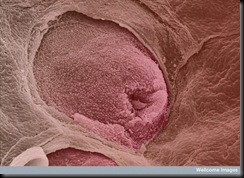
6. Tongue with taste bud

tongue This color-enhanced image depicts a taste bud on the tongue. The human tongue has about 10,000 taste buds that are involved with detecting salty, sour, bitter, sweet and savoury taste perceptions.

7. Alveoli in the lung

alevoliThis is what a colour-enhanced image of the inner surface of your lung looks like. The hollow cavities are alveoli; this is where gas exchange occurs with the blood.

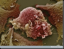
8. Lung cancer cells

lung cancer cells This image of warped lung cancer cells is in stark contrast to the healthy lung in the previous picture.

9. Villi of small intestine

villi

Villi in the small intestine increase the surface area of the gut, which helps in the absorption of food. Look closely and you’ll see some food stuck in one of the crevices.

Source: Wellcome Images